Lymphocytes

Figure 13 Types of leukocytes
These cells fall under the broader category of leukocytes, or white cells, as the separate
into the white buffy coat between red blood cells and blood plasma after
centrifugation. Lymphocytes refer to three particular agranular leukocytes, B,
T and NK cells. These cells are responsible for the destruction, identification
and proper tagging of foreign cells and possible pathogens such as bacteria or
viruses that make their way into the body. Most of these cells have a lifetime
that can be measured in weeks and must be replaced in order to maintain a
competent immune response, thus levels of cell division from lymphocyte stem
cells located in bone marrow are kept stable to provide a basal concentration
of lymphocytes in the blood. The concentration can be raised above homeostatic
norms by the presence of an infectious agent such as bacteria or viruses in
order to quickly attack and destroy the invading pathogens and mitigate the
spread of infection and any associated tissue damage. As these stem cell populations
are constantly dividing, the risk for mutation is always present and high white
cell concentrations absent of a disease state is indicative of some kind of
proliferative disorder.
B-Cells

Figure 14 3D CGI rendering of a B-cell
B-cells develop in bone marrow and then migrate to the spleen where they undergo
maturation and later release into systemic circulation where they function to
patrol the body for possible pathogens. The two main types of B-cells are memory
B-cells that have been exposed to a novel antigen during the primary immune response
and the plasma B-cells that have already been exposed to a particular antigen
and are able to secrete large amounts of antibodies specific to a particular
antigen. Once exposed to an antigen for the first time memory B-cells are able
to retain the knowledge for that specific antigen and can initiate an immune
response faster if the same pathogen is encountered again. The plasma B-cells
are clones of the memory cells and therefore each cell line will be specific to
a particular antigen and uniquely adapted to respond to repeat infection.
T-Cells

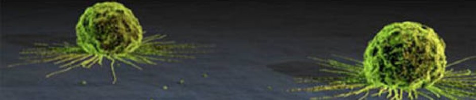
Figure 15 Colored SEM of T-Cell(pink) attacking a cancer cell(orange)
These cells also
originate in the bone marrow, but migrate to the thymus for maturation instead
of the spleen. T-cells function to destroy cells that are not recognized as
being native or “self” such as virally infected cells and allografted tissues
and organs. They are able to recognize surface proteins coded for by the major
histocompatability complex (MHC) gene loci that are present on all cells. If
the presented MHC1 sequence does not match the T-cell MHC2 sequence, the
invading cell is destroyed. Other T-cell lines are responsible for aiding other
components of the immune system by assisting in the proper identification of
pathogens and by activating B-cells once the come into contact with antigens allowing
differentiation into memory or plasma B-cells.
Natural Killer Cells (NK)

Figure 16 NK-Cell attacking intruder
The major role of these cells is to trigger apoptosis in other cells that could
potentially cause damage to tissue architecture or serious harm to the
organism. They are called natural killers as no activation is required for them
to initiate an immune response to tissue that does not present the proper MHC
proteins and will automatically release cytotoxic factors that trigger
apoptosis in the target cell. Viral cells and tumor cells that have lost proper
expression of MHC surface proteins are common targets for these cells.
To return to the overveiw of the lymph system, click here
To return to the gross anatomy of the lymph system, click here
